A major breakthrough in brain imaging promises faster, safer scans for patients and clearer insights for clinicians. By dramatically reducing scan times and revealing the brain’s blood vessel network in unprecedented detail, the research opens new opportunities for better diagnosis, monitoring, and understanding of neurological conditions.
Dr Saskia Bollmann, a Biomedical Engineer from the School of Electrical Engineering and Computer Science, is a co-author on the international study published in Science Advances.

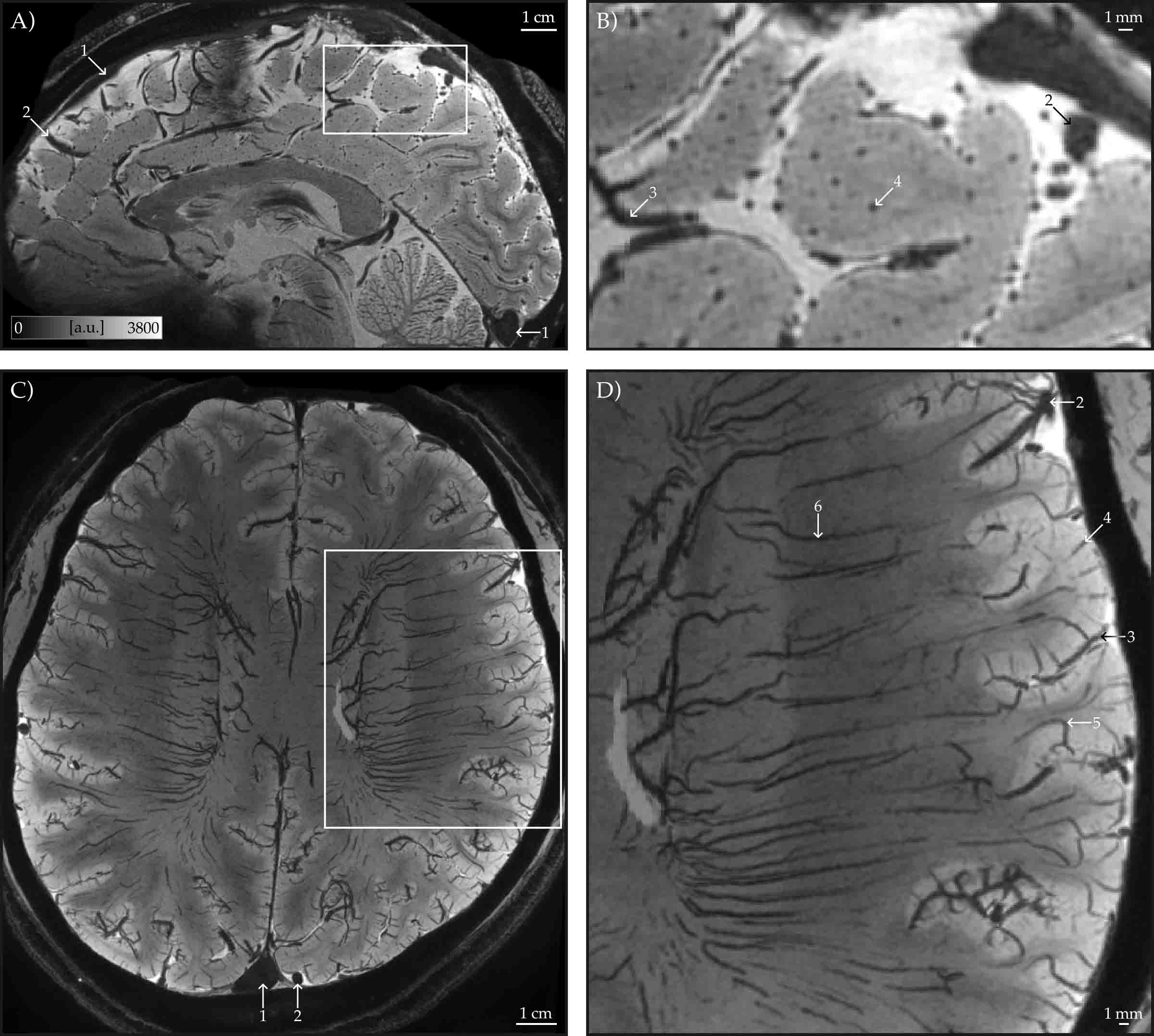
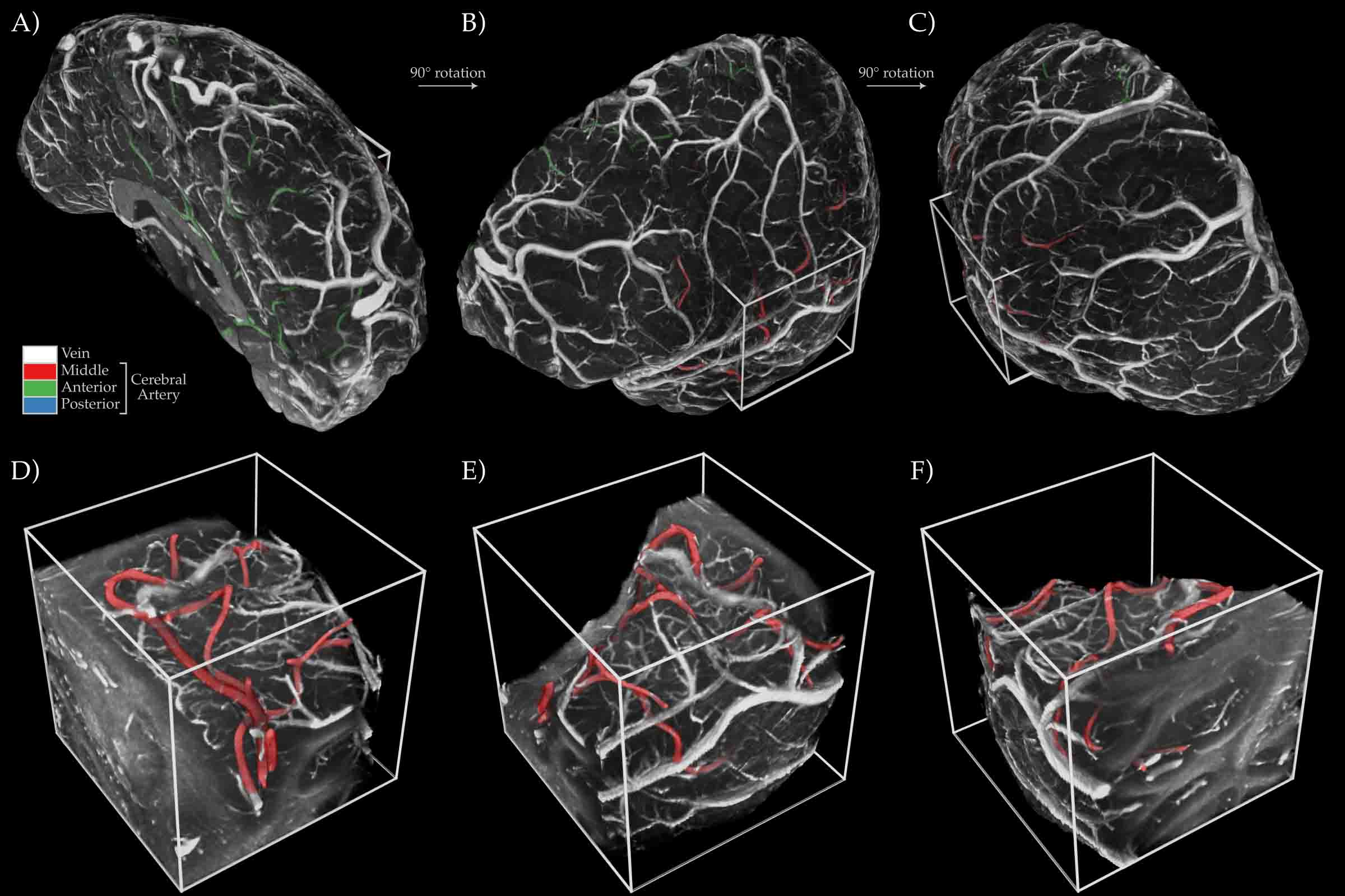
The team used ultra-high-field 7 Tesla MRI to capture whole-brain images at a resolution of 0.35 millimetres in just seven minutes, allowing tiny veins within the cerebral cortex to be visualised across the entire living brain. Blood vessels, particularly veins, strongly influence functional MRI (fMRI) signals, which are widely used to study brain activity and disease. Until now, limited visibility of the brain’s venous structure made it difficult to interpret scans accurately or detect subtle vascular changes linked to stroke, dementia, and neurodegeneration.
To overcome these challenges, the researchers developed both a rapid imaging protocol and advanced processing techniques. These methods distinguish between large surface veins, smaller pial veins, and veins embedded within the brain’s grey matter, helping clinicians separate vascular effects from true neural signals.
For patients, the faster, non-invasive scans require no contrast agents, making them more comfortable, particularly for older adults, children, and people with neurological conditions. For clinicians, detailed venous maps provide critical context for interpreting brain imaging, supporting earlier detection of abnormalities and more confident decision-making.
Dr Bollmann said, “Being able to see the brain’s tiny blood vessels in such detail and in such a short time is a major step forward. This method could really improve how we understand brain function and how vascular changes contribute to neurological disease.”
The study’s lead author, Omer Faruk Gulban, from Maastricht University highlighted the power of the team’s international collaboration.
“Breakthroughs like this are rarely the work of a single lab; they are international symphonies. While the heart of this research and the primary data collection took place at Maastricht University, the collaboration with Kendrick Kay at UMN CMRR was vital for maturing the ideas and refining our tools."

Supported by additional partners in Germany, Australia, and others in the Netherlands, this project is a testament to the power of "Team Science". It proves that when we bridge world-class expertise across borders, we can reveal the human brain in ways never before possible.”
The research also recreated classic vascular maps first produced by anatomist Henri Duvernoy and produced the first whole-brain maps of intracortical veins at this level of detail, marking a significant advance in brain imaging.
Dr Bollmann is currently based at Stanford University, where she is collaborating on research with the new 7 Tesla MRI system to further accelerate high-resolution brain imaging.
Acknowledgement: This research was conducted by an international collaboration involving Maastricht University; The University of Queensland; the University of Minnesota (Center for Magnetic Resonance Research); Harvard Medical School and Massachusetts General Hospital and Brain Innovation (Netherlands); the German Center for Neurodegenerative Diseases (DZNE); the Max Planck Institute for Empirical Aesthetics; and Friedrich Schiller University Jena.


